38 the human eye without labels
How Do We See Light? | Ask A Biologist - Arizona State University The human eye has over 100 million rod cells. Cones require a lot more light and they are used to see color. We have three types of cones: blue, green, and red. The human eye only has about 6 million cones. Many of these are packed into the fovea, a small pit in the back of the eye that helps with the sharpness or detail of images. PDF Parts of the Eye - National Institutes of Health Eye Diagram Handout Author: National Eye Health Education Program of the National Eye Institute, National Institutes of Health Subject: Handout illustrating parts of the eye Keywords: parts of the eye, eye diagram, vitreous gel, iris, cornea, pupil, lens, optic nerve, macula, retina Created Date: 12/16/2011 12:39:09 PM
60,892 Human eye anatomy Images, Stock Photos & Vectors - Shutterstock Find Human eye anatomy stock images in HD and millions of other royalty-free stock photos, illustrations and vectors in the Shutterstock collection. Thousands of new, high-quality pictures added every day.
The human eye without labels
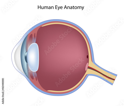
Structure and Function of the Human Eye - ThoughtCo The main parts of the human eye are the cornea, iris, pupil, aqueous humor, lens, vitreous humor, retina, and optic nerve. Light enters the eye by passing through the transparent cornea and aqueous humor. The iris controls the size of the pupil, which is the opening that allows light to enter the lens. Light is focused by the lens and goes ... The Human Eye - Diagram, Parts, Working, Function and Work of The Lens The human eye is blind for about 40 minutes every day. This is because of Saccadic masking; it is a way of the body to reduce motion blur while the object and eyes move. 20/20 is a normal vision and it's not a perfect vision. It means if a normal person can see an object at a distance of 20 feet, the test subject can also see the object at 20 feet. Category:Human eyes - Wikimedia Commons Pages in category "Human eyes" This category contains only the following page. H. Human eye; Media in category "Human eyes" The following 179 files are in this category, out of 179 total. 2016-09-21 Herstmonceux Observatorium 06.jpg. Afghani eye.png 118 × 106; 21 KB. Alfred Hitchcock's The Wrong Man trailer 02.png. An is always on you.jpg.
The human eye without labels. Structure of the Human Eye - Health Jade The eye is a hollow, spherical structure about 2.5 centimeters in diameter. Its wall has three distinct layers—an outer (fibrous) layer, a middle (vascular) layer, and an inner (nervous) layer. The spaces within the eye are filled with fluids that help maintain its shape. Figure 6. Structure of the human eye. Label Parts of the Human Ear - University of Dayton Parts of the Ear. Select the correct label for each part of the ear. Click on the Score button to see how you did. Incorrect answers will be marked in red. How the Eyes Work | National Eye Institute - National Institutes of Health How the Eyes Work. All the different parts of your eyes work together to help you see. First, light passes through the cornea (the clear front layer of the eye). The cornea is shaped like a dome and bends light to help the eye focus. Some of this light enters the eye through an opening called the pupil (PYOO-pul). diagram of eye with labels Horseshoe Crab Anatomy. 16 Pics about Horseshoe Crab Anatomy : Label the Eye, Eye With Labels Clip Art at Clker.com - vector clip art online, royalty and also Muscles of the Human Eyeball | ClipArt ETC. Horseshoe Crab Anatomy dnr.maryland.gov crab horseshoe anatomy eyes diagram labeled gills ccs dnr maryland gov
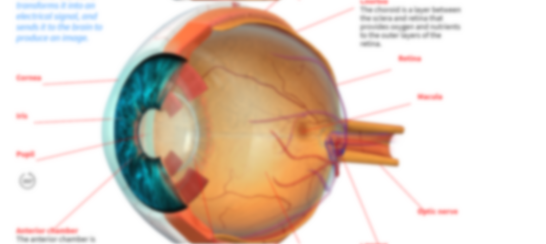
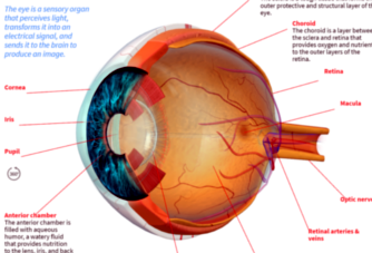
What Does the Eye Look Like? - Diagram of the Eye | Harvard Eye Associates Vitreous Gel: A thick, transparent liquid that fills the center of the eye. It is mostly water and gives the eye its form and shape. Our eyes are vital for seeing the world around us. Keep them healthy by maintaining regular vision exams. Contact Harvard Eye Associates at 949-951-2020 or harvardeye.com to schedule an appointment today. The Human Eye | Boundless Physics | | Course Hero The human eye is the gateway to one of our five senses. The human eye is an organ that reacts with light. It allows light perception, color vision and depth perception. A normal human eye can see about 10 million different colors! There are many parts of a human eye, and that is what we are going to cover in this atom. Properties ... Description Use these simple eye diagrams to help students learn about the human eye. Three differentiated worksheets are included: 1. Write the words using a word bank 2. Cut and paste the words 3. Write the words without a word bank Labels include: eyebrow, eyelid, eyelashes, pupil, iris, and sclera. Human Eye - Class 8, Light The light coming from an object enters the eye through cornea. The main function of cornea is to protect the eye but it also helps in focussing some light (by its converging action). (3) Just behind the cornea is the Iris. Iris is the coloured part of the eye. The iris has a hole at its centre which is called pupil.
The Eyes (Human Anatomy): Diagram, Optic Nerve, Iris, Cornea ... - WebMD The weaker eye, which may or may not wander, is called the "lazy eye." Astigmatism: A problem with the curve of your cornea. If you have it, your eye can't focus light onto the retina the way it... Human Eye - Definition, Structure, Function, Parts, Diagram - BYJUS Structure of Human Eye. A human eye is roughly 2.3 cm in diameter and is almost a spherical ball filled with some fluid. It consists of the following parts: Sclera: It is the outer covering, a protective tough white layer called the sclera (white part of the eye). Cornea: The front transparent part of the sclera is called the cornea. Anatomy of the eye: Quizzes and diagrams | Kenhub Here you can see all of the main structures in this area. Spend some time reviewing the name and location of each one, then try to label the eye yourself - without peeking! - using the eye diagram (blank) below. Unlabeled diagram of the eye Click below to download our free unlabeled diagram of the eye. Eye Diagram Teaching Resources | Teachers Pay Teachers The Human Eye Overview Reading Comprehension and Diagram Worksheet. by. Teaching to the Middle. 4.7. (65) $1.50. Zip. This passage briefly describes the human eye (900-1000 Lexile). 14 questions (matching and multiple choice) assess students' understanding. Students label a diagram of 6 parts of the eye.
How the Human Eye Works | Blend4Web
Human eye - Wikipedia The human eye is a sensory organ, part of the sensory nervous system, that reacts to visible light and allows us to use visual information for various purposes including seeing things, keeping our balance, and maintaining circadian rhythm . The eye can be considered as a living optical device.
The Human Eye by Second Star | Teachers Pay Teachers
Label Parts of the Human Eye - University of Dayton Parts of the Eye. Select the correct label for each part of the eye. The image is taken from above the left eye. Click on the Score button to see how you did. Incorrect answers will be marked in red. ...

Biology vs Physics: Two Ways of Doing Science? | Footnotes to Plato
Eye Diagram With Labels and detailed description - BYJUS Iris is the coloured part of the eye and controls the amount of light entering the eye by regulating the size of the pupil. The lens is located just behind the iris. Its function is to focus the light on the retina. The optic nerve transmits electrical signals from the retina to the brain. Pupil is the opening at the centre of the iris.

Human eye - Wikipedia
The Human Eye Worksheets | Human eye diagram, Parts of the ... - Pinterest Description Use these simple eye diagrams to help students learn about the human eye. Three differentiated worksheets are included: 1. Write the words using a word bank 2. Cut and paste the words 3. Write the words without a word bank Labels include: eyebrow, eyelid, eyelashes, pupil, iris, and sclera.
Activity 7 - Brain & Cranial Nerves
Eye Anatomy: A Closer Look At the Parts of the Eye - All About Vision In a number of ways, the human eye works much like a digital camera: Light is focused primarily by the cornea — the clear front surface of the eye, which acts like a camera lens. The iris of the eye functions like the diaphragm of a camera, controlling the amount of light reaching the back of the eye by automatically adjusting the size of the ...

Basic anatomy of the human eye - YouTube
Human Eye Explorer Even microscopic structures, usually not visible for the human eye, can be explored: see inside the retina or cornea and discover its layers and cells from all perspectives. Additional functions like the presentation editor, the media library or labeling and color tools, make the Human Eye Explorer a unique software.
Anatomy of the eye, non-labeled - Buy this stock illustration and explore similar illustrations ...
Eye Anatomy: Parts of the Eye and How We See Behind the anterior chamber is the eye's iris (the colored part of the eye) and the dark hole in the middle called the pupil. Muscles in the iris dilate (widen) or constrict (narrow) the pupil to control the amount of light reaching the back of the eye. Directly behind the pupil sits the lens. The lens focuses light toward the back of the eye.
How the Human Eye Works | Blend4Web
File:Diagram of human eye without labels.svg - Wikimedia Size of this PNG preview of this SVG file: 410 × 430 pixels. Other resolutions: 229 × 240 pixels | 458 × 480 pixels | 732 × 768 pixels | 976 × 1,024 pixels | 1,953 × 2,048 pixels. Original file (SVG file, nominally 410 × 430 pixels, file size: 277 KB) File information. Structured data.

Relapsing Polychondritis: Causes, Picture, Symptoms and Treatment
Quiz: Label The Parts Of The Eye - ProProfs Quiz Quiz: Label The Parts Of The Eye. Do you know the anatomy of the human eye very well? Can you label the parts of the eye in the quiz below? Give it a try and evaluate yourself. The eye has many important parts, each with different functions, including the cornea, pupil, sclera, and many more. Can you tell where these parts are located and what ...

Anatomy of the Human Eye - News-Medical.net The light passing through cornea, pupil, and lens gets focused on the retinal membrane. In addition to tissue components, retina is made up of two types of cells: rod cells and cone cells. The ...

zuhrotun nisfah anak kebumen
Eye Anatomy: 16 Parts of the Eye & Their Functions - Vision Center The lens of the eye (or crystalline lens) is the transparent lentil-shaped structure inside your eye. This is the natural lens. It is located behind the iris and to the front of the vitreous humor (vitreous body). The vitreous humor is a clear, colorless, gelatinous mass that fills the gap between the lens and the retina in the eye.
Human Skeleton Back No Text No Color Clip Art at Clker.com - vector clip art online, royalty ...
Category:Human eyes - Wikimedia Commons Pages in category "Human eyes" This category contains only the following page. H. Human eye; Media in category "Human eyes" The following 179 files are in this category, out of 179 total. 2016-09-21 Herstmonceux Observatorium 06.jpg. Afghani eye.png 118 × 106; 21 KB. Alfred Hitchcock's The Wrong Man trailer 02.png. An is always on you.jpg.

Eye Project for science: Eye Project for science
The Human Eye - Diagram, Parts, Working, Function and Work of The Lens The human eye is blind for about 40 minutes every day. This is because of Saccadic masking; it is a way of the body to reduce motion blur while the object and eyes move. 20/20 is a normal vision and it's not a perfect vision. It means if a normal person can see an object at a distance of 20 feet, the test subject can also see the object at 20 feet.

What Does The Eye Really See?
Structure and Function of the Human Eye - ThoughtCo The main parts of the human eye are the cornea, iris, pupil, aqueous humor, lens, vitreous humor, retina, and optic nerve. Light enters the eye by passing through the transparent cornea and aqueous humor. The iris controls the size of the pupil, which is the opening that allows light to enter the lens. Light is focused by the lens and goes ...
Ear | ClipArt ETC

human-eye1 | DIY Projects


Learning Chart Our Amazing Eye | Cool eyes, Human body science, Learning
Post a Comment for "38 the human eye without labels"