43 brain mri with labels
› doi › 10Sex beyond the genitalia: The human brain mosaic | PNAS Nov 30, 2015 · Previous criticisms of the dichotomous view of human brain have focused on the fact that most sex/gender differences are nondimorphic population-level differences with extensive overlap of the distributions of females and males and have therefore claimed that human brains cannot be sorted into two distinct classes: “male brains” and “female brains” (6–8). New machine learning model flags abnormal brain scans in real-time "Having previously built and validated a labeled head MRI dataset using cutting edge machine learning methodology through a team of data scientists and hospital radiologists, the same team have now...
Semisupervised Training of a Brain MRI Tumor Detection Model Using ... The application of semisupervised learning to mined image annotations significantly improved tumor detection performance, achieving an excellent F 1 score of 0.954. This development pipeline can be extended for other imaging modalities, repurposing unused data silos to potentially enable automated tumor detection across radiologic modalities.
Brain mri with labels
› pmc › articlesThe Human Brainnetome Atlas: A New Brain Atlas Based on ... Jul 25, 2016 · The Brainnetome Atlas Viewer was coded in MATLAB so that it can easily be implemented into commonly used brain MRI processing pipelines (Fig. 6 C). In addition, the atlas will be useful for the definition of masks for seeding specific a priori cortical regions or networks of interest in prospective neuroimaging studies. Ventricles of the brain: Anatomy and pathology | Kenhub This space is therefore occupied by a clear fluid that suspends the brain within the cranial vault. The fluid (cerebrospinal fluid) is produced in the ventricular system of the brain. There are four such hollow spaces in the brain that house cerebrospinal fluid (CSF): two lateral ventricles, a third ventricle and a fourth ventricle. Key facts. en.wikipedia.org › wiki › Molecular_imagingMolecular imaging - Wikipedia MRI has the advantages of having very high spatial resolution and is very adept at morphological imaging and functional imaging. MRI does have several disadvantages though. First, MRI has a sensitivity of around 10 −3 mol/L to 10 −5 mol/L which, compared to other types of imaging, can be very limiting. This problem stems from the fact that ...
Brain mri with labels. Brain Tumor MRI Dataset | Kaggle Br35H This dataset contains 7022 images of human brain MRI images which are classified into 4 classes: glioma - meningioma - no tumor and pituitary. no tumor class images were taken from the Br35H dataset. Automated MRI image labelling processes 100,000 brain exams in under 30 ... Researchers from the School of Biomedical Engineering & Imaging Sciences at King's College London have automated brain MRI image labeling, needed to teach machine learning image recognition models,... Cross-sectional anatomy of the brain - e-Anatomy - IMAIOS We created a brain atlas that is an interactive tool for studying the conventional anatomy of the normal brain based on a magnetic resonance imaging exam of the axial brain. Anatomical structures and specific areas are visible as interactive labeled images. Cross sectional anatomy: MRI of the brain. An MRI was performed on a healthy subject ... Researchers automate brain MRI image labeling, more than 100,000 exams ... Researchers have automated brain MRI image labeling, needed to teach machine learning image recognition models, by deriving important labels from radiology reports and accurately assigning them to...
Labeled imaging anatomy cases | Radiology Reference Article ... This article lists a series of labeled imaging anatomy cases by body region and modality. Brain CT head: non-contrast axial CT head: non-contrast coronal CT head: non-contrast sagittal CT head: angiogram axial CT head: angiogram coronal CT... Deep learning to automate the labelling of head MRI datasets for ... manually labelling mri scans appears to be particularly laborious due to (1) the superior soft-tissue contrast of mri which enables more refined diagnoses compared with other imaging modalities such as computed tomography; and (2) the use of multiple, complementary imaging sequences so that a larger number of images must be scrutinised per … › Lower-Limb › Ankle-Foot-MRIAnatomy of the foot and ankle - MRI - e-Anatomy - IMAIOS Sep 13, 2021 · Cross-sectional anatomy: MRI of the ankle and feet. A magnetic resonance imaging (MRI) was performed on a normal subject; with spin-echo T1 weighted images and spin-echo fat-saturated proton density weighted images (3 usual planes used for osteo-articular imaging: axial, coronal, and sagittal). Scientists automate brain MRI image labeling, more than 100,000 exams ... Researchers from the School of Biomedical Engineering & Imaging Sciences at King's College London have automated brain MRI image labelling, needed to teach machine learning image recognition models, by deriving important labels from radiology reports and accurately assigning them to the corresponding MRI examinations.
Potentially life-saving study could cut labelling times for MRI Scans ... A groundbreaking deep learning study can label 100,000 MRI scans for cancer and neuro patients in minutes rather than years. ... researchers at King's College in London have made a breakthrough ... A unified 3D map of microscopic architecture and MRI of the human brain The inclusion of five microscopy labels, blockface images, and three quantitative MRI contrasts provides a wealth of anatomical information ( Fig. 1 ). The full-brain coverage allows for detailed and comparative analyses of architectonic features for mapping the cortical laminar structure ( 20 - 23 ). New MRI probe can reveal more of the brain's inner workings Traditional fMRI imaging measures changes to blood flow in the brain, as a proxy for neural activity. When neurons receive signals from other neurons, it triggers an influx of calcium, which causes a diffusible gas called nitric oxide to be released. Nitric oxide acts in part as a vasodilator that increases blood flow to the area. data-flair.training › blogs › braBrain Tumor Classification using Machine Learning - DataFlair One such application of deep learning to detect brain tumors from MRI scan images. About Brain Tumor Classification Project. In this machine learning project, we build a classifier to detect the brain tumor (if any) from the MRI scan images. By now it is evident that this is a binary classification problem.
Magnetic Resonance Imaging (MRI): Brain (for Parents) - Nemours Kidshealth
da.si.washington.edu › daDigital Anatomist Interactive Atlases - University of Washington Jun 12, 2018 · It is organized into functional chapters suitable as a laboratory guide, with an instructive caption accompanying each image. It contains 3-D computer graphic reconstructions of brain material; MRI scans; tissue sections, some enhanced with pathways; gross brain specimens and dissections; and summary drawings.

Brain Differences in College Aged Occasional Drug Users - Neuroscience News
› Upper-Limb › Upper-extremity-MRIArm, forearm, and hand: MRI of anatomy - e-Anatomy - IMAIOS Sep 13, 2021 · Anatomy of the arm, forearm, wrist, shoulder and hand: how to view the anatomical labels. This module is a comprehensive and affordable learning tool for medical students and residents and especially for rheumatologists, orthopedic surgeons and radiologists.
Annotated Sagittal T1 Midline MRI Scan of Reigh's Brain | Flickr
› AANLIB › casesHarvard University Show labels Show list All modalities to: MR-T1 MR-T2 FDG T1/FDG T2/FDG
Magnetic Resonance Imaging (MRI) – RadLink
clara_pt_brain_mri_segmentation | NVIDIA NGC The model is trained to segment 3 nested subregions of primary brain tumors (gliomas): the "enhancing tumor" (ET), the "tumor core" (TC), the "whole tumor" (WT) based on 4 aligned input MRI scans (T1c, T1, T2, FLAIR). The ET is described by areas that show hyper intensity in T1c when compared to T1, but also when compared to "healthy" white ...

BIOLOGY BLOG, : July 2012
Segmentation Labels and Radiomic Features for the Pre-operative Scans ... this data container describes both computer-aided and manually-corrected segmentation labels for the pre-operative multi-institutional scans of the cancer genome atlas (tcga) low grade glioma (lgg) collection, publicly available in the cancer imaging archive (tcia), coupled with a rich panel of radiomic features along with their corresponding …

MRI Brain Planning
The Basics of MRI Interpretation | Radiology | Geeky Medics An overview of magnetic resonance imaging (MRI), including different sequence types (T1, T2, STIR, FLAIR) and a structured approach to MRI interpretation. ... Normal brain MR shows differences between T1 and T2 images. Licence: . Andrew Murphy, et al. MRI sequences (overview). Radiopaedia.org, the wiki-based collaborative Radiology resource.

MRI BLOG: Brachial Plexus MRI (II/II)
CoordinateSystems - Brainstorm The Brainstorm function mri_normalize_maff.m is based on SPM12 function spm_maff8.m, described in (Ashburner 2005). It is integrated in Brainstorm and does not require the installation of SPM12. This transformation is fast to compute and to apply to 3D coordinates, and very light in terms of storage.

Cross Sectional Anatomy of the Brain - Cerebral Artery - GUWS Medical
Arterial spin labeling MR perfusion - Radiopaedia Arterial spin labeling (ASL) MR perfusion is an MR perfusion technique which does not require intravenous administration of contrast (unlike DSC perfusion and DCE perfusion ). Instead it exploits the ability of MRI to magnetically label arterial blood below the imaging slab. The parameter most commonly derived is cerebral blood flow (CBF).

Dr Balaji Anvekar FRCR: Frontal subcortical white matter cystic lesions MRI
Tutorials/LabelFreeSurfer - Brainstorm Display the cortex surface on top of the MRI slices, to make sure that they are well aligned, that the surface follows well the folds, and that left and right were not flipped: right-click on the low-resolution cortex > MRI registration > Check MRI/surface registration... The cortex looks bad

MRI Musculo-Skeletal Section: How to locate glenohumeral ligaments.
fastMRI+, Clinical pathology annotations for knee and brain fully ... in this paper, we present wide availability of a complementary dataset of annotations, fastmri+, consisting of human subspecialist expert clinical bounding box labelled pathology annotations for...
Neuroscience And The Law | WUNC
Researchers automate brain MRI image labelling, more than 100,000 exams ... Now, more than 100,00 MRI examinations can be labelled in less than half an hour. Published in European Radiology, this is the first study allowing researchers to label complex MRI image datasets at scale. The researchers say it would take years to manually perform labelling of more than 100,000 MRI examinations.

MRI Procedure of Brain
Classification of brain tumours in MR images using deep ... - Nature Glioma tumours are the result of glial cell mutations resulting in malignancy of normal cells. They are the most common types of Astrocytomas (tumour of the brain or spinal cord), account for 30%...

Oil is For ...: October 2008
› pmc › articlesPerfusion MRI: The Five Most Frequently Asked Technical ... Perfusion MRI is a promising tool in assessing stroke, brain tumors, and patients with neurodegenerative diseases. Most of the impediments that have limited the use of perfusion MRI can be overcome to allow integration of these methods into modern neuroimaging protocols.

A Novel Approach for Brain Tumor Detection Using MRI Images
MRI has been used to reveal epigenetic changes in brain for first time A new form of magnetic resonance imaging can reveal where so-called epigenetic changes have occurred in the brain. The technique, which requires a special diet, can image chemical labels added to ...
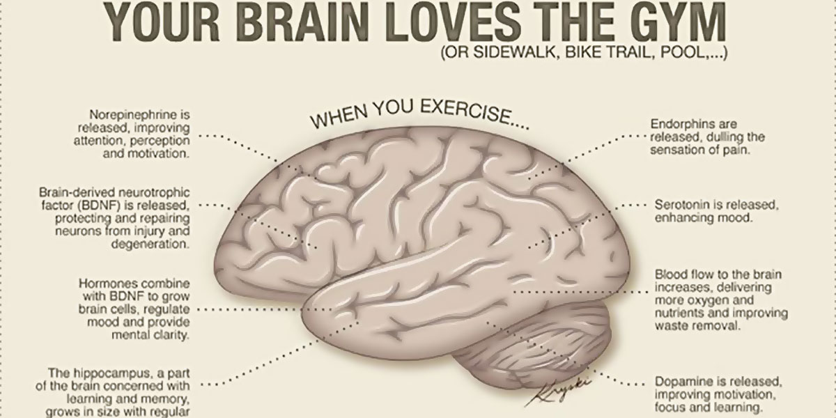
Exercise Can Help Increase Size Of The Brain’s Hippocampus
Brain: Atlas of human anatomy with MRI - e-Anatomy - IMAIOS MRI Atlas of the Brain. This page presents a comprehensive series of labeled axial, sagittal and coronal images from a normal human brain magnetic resonance imaging exam. This MRI brain cross-sectional anatomy tool serves as a reference atlas to guide radiologists and researchers in the accurate identification of the brain structures.

Brain MRI - Neurology Photo (6815446) - Fanpop
Brain Tumor/Mass Classification Framework Using Magnetic-Resonance ... These brain MRI images were collected at two hospitals in China (Nanfang Hospital and General Hospital). It had 3064 brain MRI images (1426 glioma tumors, 708 meningioma tumors, and 930 pituitary tumors); this dataset was named a dataset-III in this work. Each type of brain MRI image from all datasets is shown in Table 1. Table 1
Some sample MRI images
Brain MRI: How to read MRI brain scan | Kenhub MRI is the most sensitive imaging method when it comes to examining the structure of the brain and spinal cord. It works by exciting the tissue hydrogen protons, which in turn emit electromagnetic signals back to the MRI machine. The MRI machine detects their intensity and translates it into a gray-scale MRI image.
Post a Comment for "43 brain mri with labels"